Literature reviews on vascular dementia tend to have an “arterio-centric” view of vascular roles in disease progression. Indeed, atheroclerosis, arteriolosclerosis and cerebral amyloid angiopathy affect brain arteries and arterioles, and are associated hallmarks of cerebrovascular disease including microinfarcts, microbleeds and lacunes. However, often overlooked is that all blood flow entering the brain through arterioles must also exit through a venular system of similar caliber and organization (see image). Venules flow more slowly and are a prominent site of vascular inflammation during injury and cerebral hypoperfusion. What’s more, impaired flow in venules leads must lead to impaired upstream flow in arterioles, since everything is connected.
In this review, we dive into the details of cerebral venule network structure, and discuss the evidence that venule disease occurs in dementia. We also discuss how blockade of penetrating venules produces microinfarcts identical to that those made by occluding arterioles in rats and mice. Could venule disease in humans be an overlooked source of small-scale stroke and brain hypoperfusion? If so, venule disease should be better characterized and reported in clinical and preclinical studies. Therapeutics aimed at reducing venular pathology should also be explored in earnest.
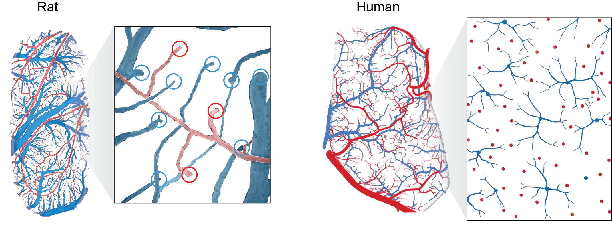
This image compares the pial arteriovenous networks of rat versus human.
Human image from Duvernoy et al, Brain Research Bulletin, 1981.